lipid rich adrenal adenoma
As in your case adrenal adenomas often are found incidentally on abdominal imaging. Lipid-rich adenomas lose signal on the chemical-shift or out-of-phase opposed-phase images while lipid-poor lesions will not lose signal.

Adrenal Adenoma Radiology Case Radiopaedia Org
An adrenal lesion with Hounsfield units of less than 10 on unenhanced CT is a benign lipid-rich adrenal adenoma.
. The lower the Hounsfield Units lipid-rich. This quantitative threshold is highly specific for the diagnosis of adenoma 7. Sharply well circumscribed appearing encapsulated Cut section. The outer cortex and the inner medulla.
Unenhanced attenuation of less than 10 HU was considered diagnostic of an adrenal adenoma. 70 of adenomas contain high intracellular fat and will be of low attenuation on unenhanced CT 45. Yellow or golden yellow with areas of dark discolouration due to hemorrhage Microscopy Appear encapsulated with pushing border having compressed fibrous capsule. Learn what causes them how to know if you might have one and how theyre treated.
Adrenal adenomas can be detected on non-contrast scans due to its low HU lipid-rich content signal drop out is seen in opp-phase. T2W diagnosis of adenoma may also be valuable in patients where chemical-shift MRI is limited due to motion artifact and in smaller nodules. In comparison to surrounding adrenal gland adenoma cells are larger with different cytoplasm increased variation in nuclear size Distinct cell borders cells have abundant foamy cytoplasm reminiscent of zona fasciculata Balloon cells. A lipid-rich mass is more likely to be a benign adenoma.
An adrenal gland adenoma is a tumor on your adrenal gland that isnt cancer but can still cause problems. Architectural pattern of cells. Thus if a lesion loses SI it is a lipid-rich adenoma Fig. Lipid-rich adenomas lose signal on the chemical-shift or out-of-phase opposed-phase images while lipid-poor lesions will not lose signal.
Attenuation measurements are performed by ROI analy-sis. If it does not lose SI all one can state is that the lesion does not contain lipid and is therefore not a lipid-rich adenoma. On MR lipid-rich adrenal adenomas may demonstrate out-of-phase signal dropout which again demonstrates that the lesion is a benign adenoma despite FDG avidity Fig. In lipid-rich adenomas atten - uation measurements will be less than 10 HU.
Its not clear what causes adrenal adenomas to form. Adrenal adenomas develop in the cortex. If it does not lose SI all one can state is that the lesion does not contain lipid and is therefore not a lipid-rich adenoma. If the mass has an attenuation value of more than 10 Hounsfield units and therefore is lipid-poor it should be removed said Dr.
The T2W properties of a lipid-rich adenoma may be useful when differentiating a lipid-rich adenoma from other adrenal masses which show microscopic fat on chemical-shift MRI including selected metastases ACC and rarely pheochromocytoma. View answer Answered by. Thus if a lesion loses SI it is a lipid-rich adenoma Fig. The absolute or relative percentage washout of contrast material on delayed contrast-enhanced CT is a highly specific test for the differentiation of lipid-poor and lipid-rich adrenal adenomas from adrenal nonadenomas.
Tumor cells have pale staining lipid rich cells with uniform round nuclei. Using a safe threshold value of 10HU on a native CT scan results in a sensitivity of 70-79 and a high specificity of 96-98 for the diagnosis of an adenoma 5-7. Primary aldosteronism Cushings syndrome. In some cases functional adrenal adenomas can be treated with medications that block the function or lower the levels of the.
Fatty are the more likely it is that the tumor is not a cancer but. Clusters of cells with enlarged lipid-rich cytoplasm seen in Cushing syndrome. They tend to be more common in older adults and people who are obese as well as in those who have diabetes or high blood pressure. The present study was undertaken to evaluate the hypothesis that lipid-rich adrenal incidentalomas a hallmark of benign adrenal adenomas may not show excess growth andor develop excess hormonal secretion during short-term follow-up and that it might be possible to re-evaluate them after 5-year follow-up instead of at 1 to 2 years intervals.
A density equal to or below 10 HU is considered diagnostic of a lipid-rich adenomas. Shailja Puri Pathologist and Microbiologist. ROIs should be placed within the nodule encompassing two-thirds of its. Removal of the affected adrenal gland usually resolves other medical conditions that may be present as a result of elevated adrenal hormones ie.
Even though the relative percentage washout of the lipid-poor adenomas was lower than that of lipid-rich adenomas it was remarkably different from that of the nonadenomas. If this would be the case it would allow for a. The lipid-rich adrenal tumors were proved to be 16 non-hormone-secreting tumors 15 adenomas and one myelolipoma and 13 hormone-secreting tumors five subclinical cortisol-producing adenomas six aldosterone-producing adenomas and two adenomas that produced both cortisol and aldosterone. Schteingart a professor of medicine and endocrinology at the University of Michigan Ann Arbor who has made particular study of incidentalomas.
I have a benign lipid rich left adrenal adenoma and a small fluid intense lesion athe left kidney. Functional adrenal adenomas are typically treated with surgery. Essentially this is a measure of how dense or fat-containing the tumor is based on measurements of what is called Hounsfield Units HU. None of the patients had pheochromocytoma or a malignant adrenal tumor.
If the initial unenhanced CT revealed adrenal nodule attenuation of less than 10 HU the nodule was considered a lipid-rich adenoma. Adrenal adenoma lipid rich or lipid poor Often homogeneous Increase in enhancement from arterial to venous phase Typically enhances 100 HU arterial 130 HU. Adrenal protocol CT was interpreted according to the following standard. Adrenal metastases should not have Hounsfield units of less than 10 on unenhanced CT.
The detection of intracytoplasmic lipid found within adrenal adenomas which reduces their CT attenuation. Each adrenal gland has two parts. Nesting alveolar trabecular. 70 pounds and still very limited on eating as well I have all the symptoms that.

Adrenal Adenoma Radiology Case Radiopaedia Org

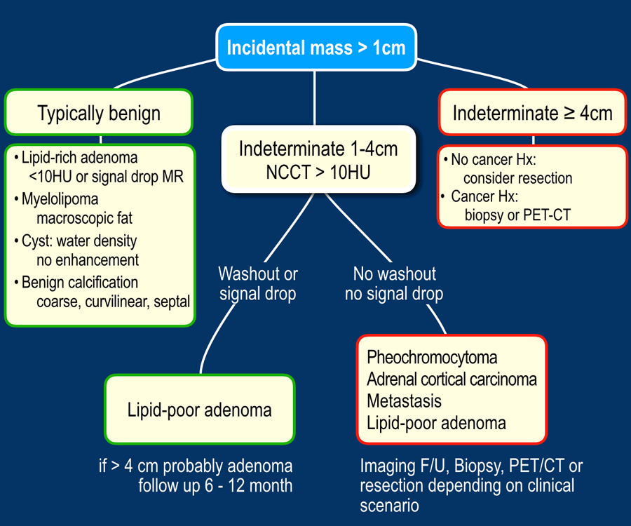
The Radiology Assistant Characterization Of Adrenal Lesions

The Radiology Assistant Characterization Of Adrenal Lesions

The Radiology Assistant Characterization Of Adrenal Lesions

Chemical Shift Mr And Precontrast Ct Scans Of Right Lipid Rich Adrenal Download Scientific Diagram

X Rays Ct Scans Mri And Other Tests For Adrenal Glands

Posting Komentar untuk "lipid rich adrenal adenoma"